TMJ/Pain

TMJ is the acronym for temporomandibular joint. It is the joint just in front of the ear canal and can be felt by placing your index finger in front of the ear canal as you open and close the lower jaw. There are 2 joints (right and left) that are part of the lower jaw (mandible). Each joint is comprised of the following structures (see figure 1 below):
Glenoid Fossa - the boney concavity of the skull that surrounds the the disc and condyle.
Condyle - the head of the joint on the mandible.
Disc - a cartilage like structure that is situated between the condyle and the glenoid fossa
Ligaments - that hold the condyle and disc to the fossa
Lateral Pterygoid Muscle - There are 2 parts the this muscle:
Lower portion (inferior head) - is responsible for pulling the head of the condyle forward
towards the midline.
Upper portion (Superior head) - is responsible for guiding the disc back to a seated joint
position upon
closing.
Pain from the TMJ area can be from 1 of 3 types and are listed from most to least common:
Muscle Pain - is most commonly from the muscles used for moving the lower
jaw forward and side to side, and the muscles to close the lower jaw and clench. Muscle
pain can be acute or chronic. Many symptoms of chronic muscle pain, tension, or
tenderness can lead to satellite pain patterns that involve pain in other muscles of the
head and neck.
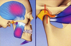
Many tension headaches can be manifested from disharmony of the bite/joint
relationship. Muscle spasm can be a trigger for a migraine to start. In addition, ringing
in the ears (tinnitus) can result from chronic muscle spasm. In many cases, it is the lateral
pterygoid muscles that are in spasm and are very sensitive to palpation. This is usually
associated with a disharmony between where the teeth fit together (maximum
intercuspation) and where the condylar head and disc want to be (see figure 2 above).
Intracapsular Pain - pain that radiates from within the joint and surrounding ligaments. This
is usually from macro-trauma directly to the lower jaw, or the the head and neck in
general with subsequent whiplash to the lower jaw and structures of the TMJ. The pain
can be acute with a recent onset within a few weeks, or can be chronic with discomfort
present for months or years (see video next page). Most commonly there is
inflammation within the joint that detected on a TMJ MRI.
Osteoarthritic Pain - pain from structural changes of the bone, most commonly bone
marrow edema and/or the condylar head and detected on a TMJ MRI or CT scan.
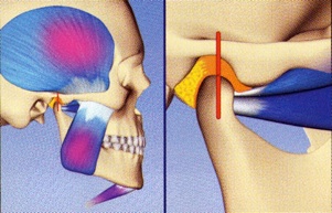
Normal seated joint position with teeth together with relaxed lateral pterygoid muscles
Forward joint position with teeth together and contracted lateral pterygoid muscles
Figure 1
Figure 2